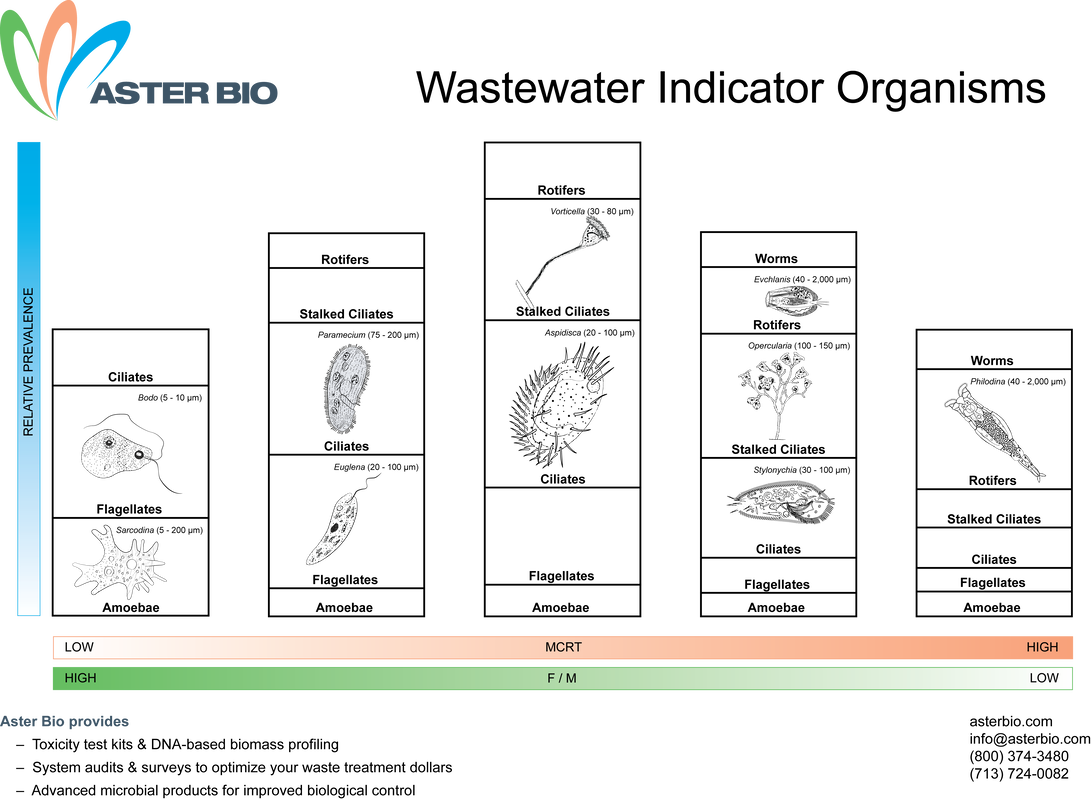
In performing a daily microscopic exam, I like to see operators noting the following:
- Note floc size and density. While phase contrast helps to see filaments, you can also see filaments with regular light microscopy. You do not need to do full filament ID with staining here. Just note floc and filaments that you see and compare your observations with SV30/SVI, turbidity, and other system data.
- Note protozoa present - you do not have to identify down to genus level! Just note amoeba, flagellates, free-swimming ciliates, crawling ciliates, and stalk ciliates. These are morphological types which only require quick observation. Also note multi-cellular organisms if present.
Now that you have the observations, you can note where the system is on the growth curve. Remember, most system are designed to function best in the early stages of decline phase growth. This region of the growth curve gives low soluble BOD5, efficient nitrification, and good floc formation - all good things.
While the bug chart says you will see a biomass dominated by stalk ciliates, crawling ciliates, and some metazoa, you may have system specific differences. This is why you want to perform frequent microscopic exams on your system to know how a good, normal indicator organism population looks. If you see changes in the population, you can look for movement along the growth curve towards "young sludge" or "old sludge" and make operational changes as needed.


 RSS Feed