• Blog
  • Lab Testing
  • Bioaugmentation Applications
  • Useful Information
  • About Us
BIOLOGICAL WASTE TREATMENT EXPERT
Contact Us

India Ink test - an easy way to monitor floc extracellular polymers

1/31/2019

 
Picture
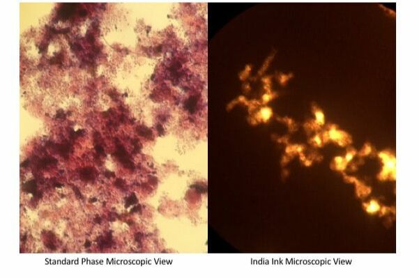
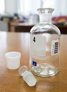
The easiest way to gauge EPS in activated sludge floc is to use the India Ink test with your existing microscope. In addition to being easy, India Ink requires no specific equipment or skills - just a bottle of $10 India Ink that will last for years. Unlike most inks, India Ink contains finely ground carbon particles that are suspended in water. You can find India Ink in many craft stores or online - Amazon India Ink with dropper. 

To use India Ink with your microscope follow these steps:
  1. Pipette a small droplet of MLSS onto a glass slide 
  2. Add an even smaller droplet of India Ink to the slide - just enough to color the water droplet dark gray/black.
  3. Place coverslip on the slide
  4. Observe using a standard microscope - you can use either regular light objective or phase contrast.
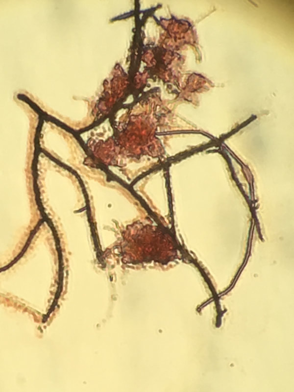
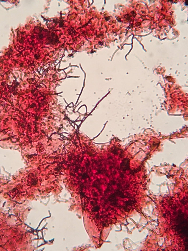
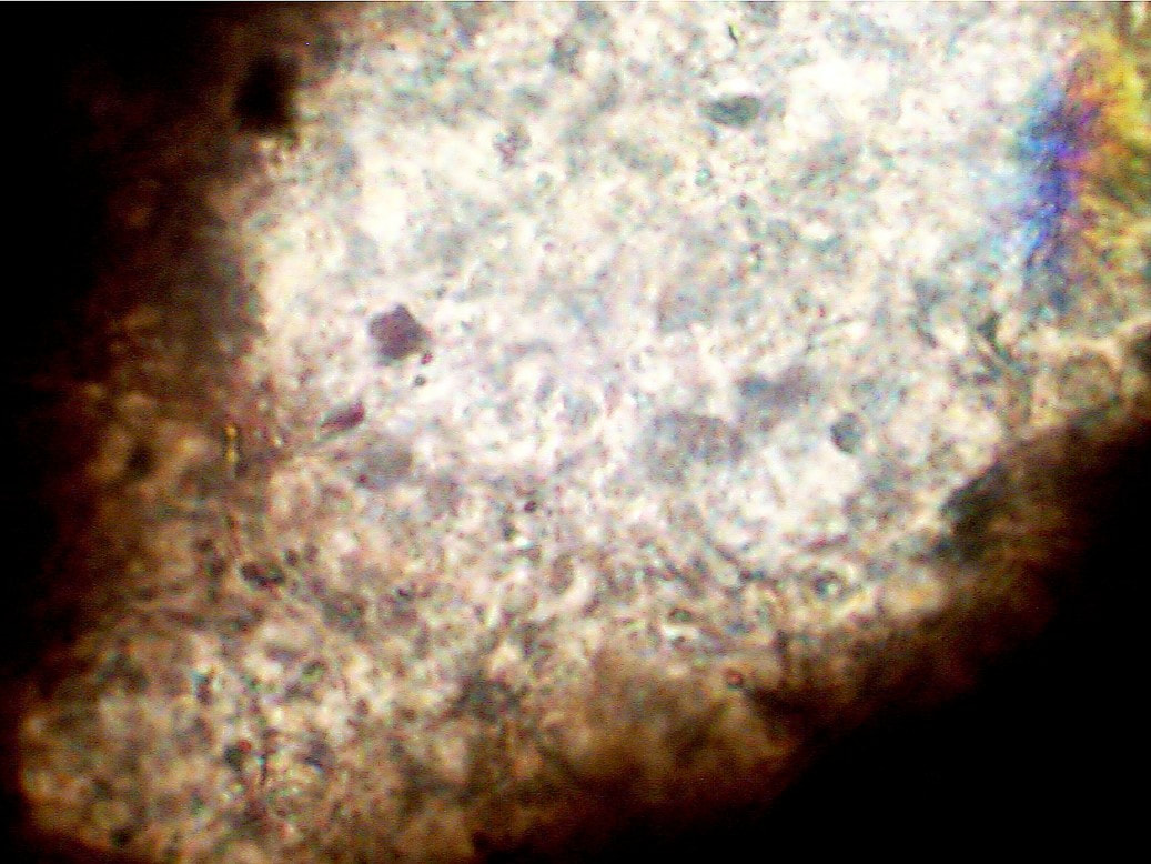
  5. The India Ink should move through the water and stain the slide. The clear zones with light shining through are floc with high levels of EPS that India Ink does not penetrate.
  6. Note the size and number of clear zones in the droplet.
  7. That is all that is required. I recommend doing India Ink tests at least 2x weekly and note any changes in EPS.
With respect to EPS, remember that some is needed to function as the glue that keeps floc together. Probelems arise when excess EPS creates a gelatin like matrix that entraps water and creates viscous bulking/non-filamentous bulking conditions.

What Oxygen Uptake Rate (OUR) test results mean for wastewater bacteria

1/28/2019

 
Picture
Standard BOD bottle used in OUR test. Photo from Wikimedia Commons.
Oxygen Uptake Rate testing is often done without much thought and we report a number, making sure it is within a specific range. Let's step back for minute and understand what the OUR number really means and how it changes in response to influent quality. 

We run OUR using simple equipment - a DO meter and BOD bottle which is present in most labs. No reagents or special techniques are needed. A PDF of the standard OUR & SOUR protocol is at the bottom of the post.
​
OUR is simply how much oxygen is consumed over a short-time period in a stirred BOD bottle. The OUR gives microbial respiration rates for the MLSS (living bacteria in the bottle). Like humans, bacteria use more oxygen when they are rapidly growing and dividing. What causes rapid growth and cell division? High levels of soluble organics that are a source of food for the microbes. So if we were to spike MLSS with sugars, we would see a rapid increase in respiration rates. A rapid decrease in OUR can be equally problematic. For instance, a shock loading of phenol, solvents, or other biocidal influent can kill many of the living organisms. There is a temporary drop in OUR due to the biocidal action. This is followed by a rapid increase in OUR as the toxicity decreases and the microbes return to steady-state populations. On the growth curve this represents low to decline phase growth where biological wastewater systems are in optimal health.
dour___sour_test.pdf
File Size: 361 kb
File Type: pdf
Download File

Using qPCR to warn of increasing Nocardia organisms in wastewater foam

1/22/2019

 
Picture
Molecular testing, including qPCR, allows for direct, rapid analysis of microbes present in a sample. We can look for important good organisms such as ammonia oxidizing bacteria, or bad organisms such as Nocardia foaming bacteria. How does it work?

Genetic material from foam samples is extracted and we identify the specific organisms responsible for Nocardia foam outbreaks. From testing, we have found most Nocardia foams are caused by a bloom of Gordonia species. We use Gordonia specific primers, qPCR gives quantitive data within a few hours of starting the test.

The qPCR advantage stems from its sensitivity and low per test cost. Unlike older microscopic or plate count techniques, qPCR can detect foaming microbes at much lower levels before they become a problem. You can adjust wasting and take corrective actions before the heavy, problem foaming.

Using Gram Stain to assist in microscopic exam of floc

1/15/2019

 
Picture
Nocardia Gram Stain - Note the Gram Positive result. (Taken with a phone camera)
Looking at filaments and floc structure is difficult with standard light microscopes. While adding phase contrast helps improve filament and floc former visibility, it is still not as effective as using stains to make cell walls jump out.

Gram staining is a common method to identify bacteria with a microscope. The stain differentiates bacteria into gram-positive or gram-negative based on their cell wall composition. Often used in medical or laboratory settings, it can also be useful in wastewater samples. You need to realize in wastewater with particulates and EPS, gram stain is often variable - so unless you have an non-floc organism such as Nocardia that stains strongly gram positive, you do not have large amounts of contrast. 

Using a Gram Stain kit is easy. I'll walk through how I use it in wastewater samples.
  1. Pipette MLSS onto a slide - approximately 0.02 ml - you want enough to stain, but avoid big clumps of MLSS.
  2. Dry the slide for at least 20 minutes at room temperature or use a heat source to speed the process. Sample must be dry before starting the stain procedure.
  3. Organize the Gram Stain Solutions - (1) Gram Violet, (2) Gram Iodine, (3) Ethanol, and (4) Gram Safranin in order. 
  4. Wear latex gloves to prevent staining your hands!
  5. Have a timer handy - once you start staining, time is important.
  6. Flood the slide with Gram Violet and allow to stain for 60 seconds.
  7. Rinse slide briefly with DI water.
  8. Flood the slide with Gram Iodine and allow to stain for 60 seconds.
  9. Rinse slide with DI water.
  10. Decolorize with the ethanol. Using a dropper, drip the ethanol over the slide drop by drop for 15 seconds. Key is to decolorize but not over-decolorize. You will get a feel for this as you perform more stains.
  11. Rinse with DI water.
  12. Flood slide with Gram Safranin and allow to stain for 60 seconds.
  13. Rinse with DI water.
  14. Allow slide to dry before examining under light microscope. Do not use the phase objectives.
Be ready to be impressed by how much detail you see with the Gram stain. Cell walls should be visible including filaments with a high level of detail. If you have Nocardia foam, the foam will contain Gram Positive (dark purple) filaments that stand out.

Picture
A gram stain of floc. Photo with a phone rather than true camera setup.

Extracellular Polymeric Substances - the "glue" binding floc & biofilms

1/10/2019

 
Picture
India Ink is used to evaluate EPS levels in floc.
As microbial cells develop into floc or biofilm, they are bound tegether by Extracellular Polymeric Substances (EPS). Consisting of a mixture of polysaccharides, proteins, enzymes, and DNA, EPS provides several benefits to the microbial community:
  • Protection from the external environment including pH swings, dehydration, predators, and toxic compounds.
  • Storage of excess organic compounds or particulate organics for later use by the microbes
  • Accumulate and store valuable macronutrients & micronutrients such as phosphate and trace metals.
While EPS is needed for floc formation, certain conditions can promote excess EPS production which can lead to non-filamentous or Zoogleal bulking. This phenomenon is seen with high levels of soluble organic acids near the influent (EPS acts to store food). We also see Zoogleal bulking with low nutrients where EPS is used to store scarce compounds such as phosphate. EPS can also be capsular which is attached to cell walls which is beneficial in most cases. Under stress conditions such as pH shock, inhibitory compounds including heavy metals, EPS can become non-capsular and create a "gel" that protects the individual cells but creates problems with solids separation.

Various "flavors" of wastewater ammonia oxidation and nitrogen removal

1/8/2019

 
Ammonia, nitrite, and nitrate removal across biological wastewater treatment systems gives many operators fits. Why? Unlike organic compound degradation, ammonia nitrogen goes through several distinct steps to detoxify and finally remove the nitrogen from the water. Today I am going to cover the various groups of microbes involved in the wastewater nitrogen cycle.
  • AOB (Ammonia Oxidizing Bacteria) - the first and possibly the most critical step is the conversion of ammonia into nitrite. Multiple organisms can do this step including: Nitrosomonas, Nitrosospira, and Nitrosococcus. 
  • NOB (Nitrite Oxidizing Bacteria) - these organisms convert the nitrite produced by the AOB into nitrate. Organisms performing this step include: Nitrobacter, Nitrococcus, Nitrospina, and Nitrospira.
  • Denitrifiers - many wastewater bacteria under anoxic conditions can use nitrate/nitrite as an alternative to oxygen in respiration. Some organisms even use nitrite and nitrate under aerobic conditions. The most active genera of denitrifiers include Pseudomonas, Paracoccus, and Hyphomicrobium.
  • COMAMMOX (Complete Ammonia Oxidation) - these microbes can do both ammonia and nitrite oxidation inside one cell. In this group we have a subset Nitrospira. 
  • ANAMMOX (Anaerobic Ammonium Oxidation) - an interesting way to save aeration energy by harnessing an interesting microbial biochemistry. Anammox systems use AOB to convert ammonia into nitrite. Then under anoxic conditions, a group of microbes can take nitrite (NO2) + ammonium (NH4) into nitrogen gas and water. These microbes have several interesting properties and have an extremely slow growth rate. Implementing Anammox technology often includes running a long sludge age and incorporation of media which allows for Anammox cultures to remain fixed in biofilm. 
  • Heterotrophic Nitrification - organisms including Pseudomonas and Paracoccus species have demonstrated ammonia oxidation pathways while growing on organic compounds. In addition to being denitrifiers, these strains also oxidized ammonium. Factors and environments that encourage this process merit further study and may result in new ways to meet ammonia/nitrite treatment goals.

    Author

    Erik Rumbaugh has been involved in biological waste treatment for over 20 years. He has worked with industrial and municipal wastewater  facilities to ensure optimal performance of their treatment systems. He is a founder of Aster Bio (www.asterbio.com) specializing in biological waste treatment.

    View my profile on LinkedIn

    RSS Feed

    Click to set custom HTML

    Archives

    May 2025
    April 2025
    March 2025
    February 2025
    January 2025
    December 2024
    November 2024
    October 2024
    April 2024
    March 2024
    February 2024
    December 2023
    September 2023
    August 2023
    July 2023
    June 2023
    May 2023
    April 2023
    February 2023
    January 2023
    December 2022
    November 2022
    October 2022
    September 2022
    August 2022
    June 2022
    May 2022
    March 2022
    February 2022
    January 2022
    November 2021
    October 2021
    September 2021
    August 2021
    June 2021
    April 2021
    March 2021
    February 2021
    December 2020
    November 2020
    October 2020
    September 2020
    August 2020
    July 2020
    June 2020
    May 2020
    April 2020
    March 2020
    February 2020
    January 2020
    December 2019
    November 2019
    October 2019
    September 2019
    August 2019
    July 2019
    June 2019
    May 2019
    April 2019
    March 2019
    February 2019
    January 2019
    December 2018
    November 2018
    October 2018
    September 2018
    August 2018
    July 2018
    June 2018
    May 2018
    April 2018
    March 2018
    February 2018
    January 2018
    December 2017
    November 2017
    October 2017
    September 2017
    August 2017
    July 2017
    June 2017
    May 2017
    April 2017
    March 2017
    February 2017
    January 2017
    December 2016
    November 2016
    October 2016
    September 2016
    August 2016
    July 2016
    June 2016
    May 2016
    April 2016
    March 2016
    February 2016
    January 2016
    December 2015
    November 2015
    October 2015
    September 2015
    August 2015
    July 2015
    June 2015
    May 2015
    April 2015
    March 2015
    February 2015
    January 2015
    December 2014
    November 2014
    October 2014
    September 2014
    August 2014
    July 2014
    June 2014
    May 2014
    April 2014
    March 2014
    February 2014

Proudly powered by Weebly
Photos from Picturepest, marcoverch, perzonseowebbyra, Picturepest, Picturepest, dsearls, dungodung, Massachusetts Office of Travel & Tourism, aqua.mech, vastateparksstaff, hile, Aaron Volkening, amishsteve, Neil DeMaster, mklwong88, KOMUnews, Picturepest, kaibara87, Bernd Thaller